2005 IEEE International Conference on Acoustics, Speech, and Signal Processing
March 18-23, 2005 • Pennsylvania Convention Center/Marriott Hotel • Philadelphia, PA, USA
ICASSP
30th Anniversary
Tutorial TUT-11: Signal and image processing issues in molecular and cellular imaging
Instructors
R. Murphy; Carnegie Mellon University
Time & Location
Saturday, March 19, 13:30 - 16:30, Location: CC: Room 111
Abstract
This tutorial focuses on evolving signal processing applications and questions derived from the growing systematic use of fluorescence microscope imaging of cells and the molecules within them, and more specifically on issues relating to the new field of “location proteomics.” The field of proteomics tries to reveal how proteins work in cells by studying all proteins in parallel using high-throughput methods, and location proteomics deals specifically with automated, sensitive and objective determination of the locations of all proteins within cells.
The Human Genome Project and other genome projects have led to the population of large sequence databases as well as methods such as BLAST for retrieving sequences from them. The focus of most biomedical research is now shifting from simply identifying gene sequences to determining the properties and functions of the proteins encoded by those genes. Since mammalian cells have a number of distinct subcompartments (such as the nucleus, mitochondria, and lysosomes) that play different roles, knowledge of the subcellular locations of all proteins is critical to understanding their functions and the mechanisms by which those functions are accomplished. Using fluorescence microscopy, we can illuminate and track the molecules of a given protein and take a photograph of them inside a human cell; this way we can figure out the places in the cell where a protein is found.
Current approaches to using fluorescence microscopy rely on visual interpretation of images and description of proteins with a limited vocabulary. Recent work provides an automated and systematic alternative that involves creation of databases of fluorescence microscope images for many different proteins and tools for automated analysis and retrieval of images from those databases. The most important results from this work are that the automated systems are capable of distinguishing all major subcellular patterns with high precision and recall (>98%) and that they can resolve the patterns of proteins that were indistinguishable by visual examination.
The combination of these automated methods with methods for large-scale fluorescent tagging of proteins (such as CD-tagging) and automated fluorescence microscopes enables the new field of Location Proteomics, providing for the first time sensitive, objective and systematic determination of the subcellular location for all proteins in a proteome. This approach also permits determination of the ways in which these locations change in response to a variety of phenoma, including growth and development, onset of disease, or exposure to drugs or pathogens.
Outline:
- problem definition and introduction to relevant cell and molecular biology
- review of relevant principles of fluorescence microscopy
- current approaches to feature extraction from cell images
- results on supervised and unsupervised learning on microscope images
- current approaches to creating databases of fluorescence microscope images
- automated retrieval of images from on-line journal articles
Target Audience: Engineers and computer scientists interested in learning about the challenges and opportunities in this new area of computational biology research, as well as current methods for interpretation and retrieval of complex images that differ dramatically from natural scenes.
Presenter Information

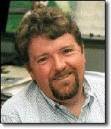
Robert F. Murphy earned an A. B. in Biochemistry from Columbia College in 1974 and a Ph.D. in Biochemistry from the California Institute of Technology in 1980. He was a Damon Runyon-Walter Winchell Cancer Foundation postdoctoral fellow at Columbia University from 1979 through 1983, after which he became an Assistant Professor of Biological Sciences at Carnegie Mellon University. He received a Presidential Young Investigator Award from the National Science Foundation shortly after joining the faculty at Carnegie Mellon in 1983 and has received research grants from the National Institutes of Health, the National Science Foundation, the American Cancer Society, the American Heart Association, the Arthritis Foundation, and the Rockefeller Brothers Fund. He has co-edited two books and published over 100 research papers. His research group at Carnegie Mellon focuses primarily on the application of fluorescence methods to problems in cell biology, with particular emphasis on automated interpretation of fluorescence microscope images. He has a long-standing interest in computer applications in biology, and developed the first formal undergraduate degree program in computational biology in 1987. He also founded the Merck Computational Biology and Chemistry Program at Carnegie Mellon in 1999. In 1984, he co-developed the Flow Cytometry Standard data file format used throughout the cytometry industry and he is Chair of the Cytometry Development Workshop held each year in Asilomar, California. He is currently Professor of Biological Sciences and Biomedical Engineering, Voting Member in the Center for Automated Learning and Discovery in the School of Computer Science, and Director of the Center for Bioimage Informatics at Carnegie Mellon.